Basic Heart Development Timeline

Embryonic Folding
The disc-like embryo then undergoes a process of folding, in which both the cranial and lateral parts of the embryo fold ventrally (forwards). This brings the heart-forming region to a ventral (frontal) position. The following animation shows the development of the heart tubes and how embryonic folding brings them to fuse in the midline.
[ads2]Segments of the Heart Tube

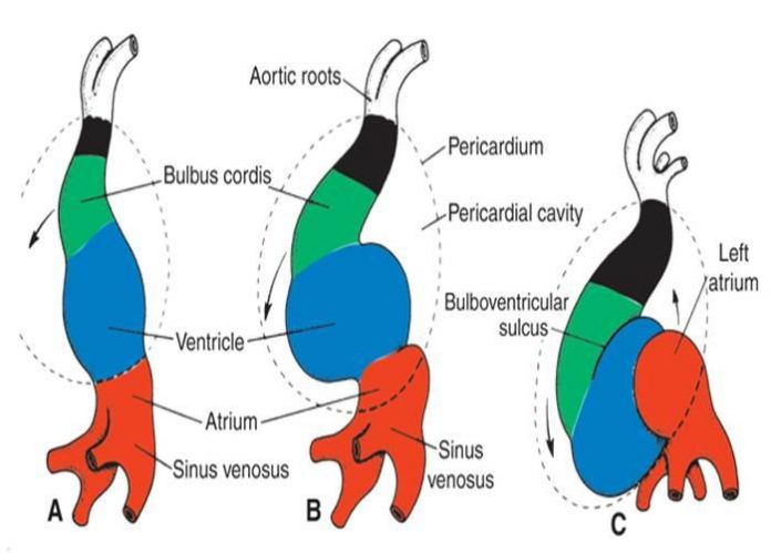
At this stage the tube already has minor constrictions within it, indicating sections of the heart tube that will form parts of the adult heart. The most caudal (tail end) segment of the heart tube is the sinus venosus which will later become the ends of the major veins carrying blood to the heart as well as parts of the atria. The next segments are the primitive atrium and primitive ventricle which will become the atria and ventricles of the adult heart. Cranial to these segments are the bulbus cordis, most of which will become the right ventricle, and the truncus arteriosus which forms the pulmonary and aortic trunks carrying blood away from the heart.
Heart Tube Looping
This tubular heart undergoes a process of looping during week four of development to form a shape that resembles that of the adult heart. It initially forms a C-shape (with the convex portion of the C situated on the right side of the embryo) and then an S-shape. Eventually the atria are brought backwards and upwards so that they lie cranially and behind the ventricles.
- The heart tube undergoes dextral looping (bends to the right) and rotation.
- The upper truncus arteriosus (ventricular) end of the tube grows more rapidly and folds downward and ventrally and to the right.
- The atria and sinus venosus lower part of the tube fold upward and dorsally and to the left. these folding begins to place the chambers of the heart in their postnatal anatomic positions.
[highlight color=”green”]The following animation outlines this process.[/highlight]
[ads2]
Septation of the Heart
The internal heart then undergoes significant changes in order to form the atria and ventricles of the adult heart. These can be summarised as follows:
| 1. Cells from the dorsal and ventral (back and front) walls of the heart grow and form two protrusions called theendocardial cushions. These grow towards each other and fuse to form the left and right atrioventricular canals. | 2. Within the primordial atrium a septum (the septum primum) grows towards the endocardial cushions. The space between the cushions and septum is known as the foramen primum. As the foramen primum decreases in size a second opening forms in the septum: theforamen secundum. | ||
| 3. A second septum (septum secundum) develops on the right of the septum primum. | 4. A primordial muscular ridge exists in the floor of the ventricle. As the left and right ventricles grow, their medial (midline) walls fuse to form the interventricular septum. | ||
| 5. Within the bulbus cordis andtruncus arteriosus, which form theoutflow tract, small ridges develop. They are continuous throughout the outflow tract and form a spiral shape. | 6. As these ridges fuse they create a spiral shaped septum throughout the outflow tract. The original outflow tract is therefore separated into both the aortaand pulmonary trunk. |
All of the partitioning of the primitive heart occurs between the middle of the fourth week and the end of the fifth week. The following animation shows the processes involved in the division of the atrioventricular canal, atria and ventricles.
| [ads1] | [box type=”note” align=”alignleft” class=”” width=”600px”]Thus the heart begins to resemble the adult heart in that it has two atria, two ventricles and the aorta forming a connection with the left ventricle while the pulmonary trunk forms a connection with the right ventricle.[/box] |
Vascular Heart Connections
Some understanding of the vascular system of an embryo is useful in completely understanding cardiac development. In the primitive heart tube section it was stated that the blood islands dispersed throughout the embryo form the early blood vessels. The islands coalesce to form vessels and these expand and continue to develop, forming vascular networks. The vascular system can be thought of in terms of arteries and veins and the major embryonic vessels can be seen in the diagram on the right.
Development of Veins
We already learnt that blood travels through the embryonic heart from the sinus venosus. There are three paired veins which form to drain into the sinus venosus:
- Vitelline veins – return poorly oxygenated blood from the yolk sac

Embryonic circulation - Umbilical veins – carry well-oxygenated blood from the primordial placenta
- Common cardinal veins – return poorly oxygenated blood from the body of the embryo
The sinus venosus soon shifts to the right to be incorporated into the right atrium as there is a shift in the venous system from the left to the right side of the embryo. The inferior vena cava and superior vena cava form and drain into the sinus venosus. In the left atrium the four pulmonary veins form, which will return oxygenated blood from the lungs.
Development of Arteries
We saw in the fusion animation that the dorsal aortae develop at the same time as the early heart tubes. These connect to the heart tubes prior to fusion via the firstaortic arch arteries. Other arches develop, which go on to form the arteries of the head and neck. We also previously saw the way in which the aorta and pulmonary trunk form. The dorsal aorta gives off branches which supply blood to the rest of the embryo:
- Gut (ventral/front) branches
- Lateral (side) branches
- Intersegmental arteries
Fetal Circulation
As the embryo progresses to a fetus the vasculature is still remarkably different to that of the adult, including the presence of three vascular shunts:
- foramen ovale (seen previously) – blood travels from the right atrium to the left atrium
- ductus venosus – blood from the umbilical vein bypasses the liver to enter the inferior vena cava
- ductus arteriosus – blood passes from the pulmonary trunk into the aorta
These shunts allow blood to bypass the lungs, liver and kidneys, whose functions are performed by the placenta while in utero.
The following diagram shows the movement of blood throughout the fetal circulation. The main flow of blood is as follows:
- [highlight color=”blue”]Placenta → umbilical vein → ductus venosus → inferior vena cava → right atrium → foramen ovale → left atrium → left ventricle → aorta → hypogastric arteries → umbilical arteries → placenta.[/highlight]
Blood that passes from the right atrium to the right ventricle travels:
- [highlight color=”blue”]Right ventricle → pulmonary trunk → ductus arteriosus → aorta[/highlight]


